Bacteriophage/Citable Version
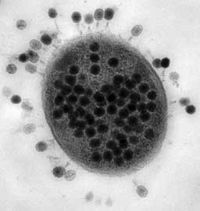
A bacteriophage (from 'bacteria' and Greek φαγειν, 'to eat') is a virus that infects bacteria. The term is commonly used in its shortened form, phage. Phages are ubiquitous and can be found in all habitats populated by bacterial hosts, such as soil or the intestine of animals. One of the densest natural sources for phages and other viruses is sea water, where up to 106 phages per ml (or, at least, of virus-like particles)(Wommack & Colwell 2000). Whitman et al. (1998) argue that there are between 1030 and 1031 prokaryotic cells on our planet. If we assume numerically one virus for every prokaryote host, then we conservatively (e.g., Bergh et al. 1989) reach a total worldwide abundance of 1030 to 1032 virus-like particles.
History
In 1896, M. E. Hankin reported that something in the waters of the Ganges and Jumna rivers in India had marked antibacterial action against cholera and could pass through a very fine porcelain filter. In 1915, British bacteriologist Frederick Twort, superintendent of the Brown Institution of London, discovered a small agent that infects and kills bacteria. He considered the agent either 1) a stage in the life cycle of the bacteria, 2) an enzyme produced by the bacteria itself or 3) a virus that grows on and destroys the bacteria. Twort's work was interrupted by the onset of World War I, and when he returned to the Brown Institution, he spent the rest of his career trying to grow bacteriophages on artificial medium. Independently, French-Canadian microbiologist Félix d'Hérelle, working at the Pasteur Institute in Paris, announced on September 3, 1917 that he discovered "an invisible, antagonistic microbe of the dysentery bacillus". For d’Herelle, there was no question as to the nature of his discovery: "In a flash I had understood: what caused my clear spots was in fact an invisible microbe... a virus parasitic on bacteria." D'Herelle called the virus bacteriophage (Stent 1963).
Structure
1. Size - Most phages range in size from 24-200 nm in length.
2. Head or Capsid - All phages contain a head structure which can vary in size and shape. Some are icosahedral (20 sides), others are filamentous. The head or capsid is composed of many copies of one or more different proteins. Inside the head is found the phage's genetic material (i.e. nucleic acid). The genetic material can be ssRNA, dsRNA, ssDNA, or dsDNA between 5 and 500 kilo base pairs (kbp) long in either a circular or linear arrangement.
3. Tail - Many, but not all, phages have tails attached to the phage head. The tail is a hollow tube through which the nucleic acid passes during infection. The size of the tail can vary considerably. In the more complex phages, like T4, the tail is surrounded by a contractile sheath which contracts during infection of the bacterium. At the end of the tail, some phages have a base plate and one or more tail fibers attached to it; these structures are involved in the attachment of the phage to the bacterial cell.
Classification
An alphabetical list of the bacteriophage families
Corticoviridae icosahedral capsid with lipid layer, circular supercoiled dsDNA
Cystoviridae enveloped, icosahedral capsid, lipids, three molecules of linear dsRNA
Fuselloviridae pleomorphic, envelope, lipids, no capsid, circular supercoiled dsDNA
Inoviridae genus Inovirus long filaments with helical symmetry, circular ssDNA
Inoviridae genus Plectrovirus short rods with helical symmetry, circular ssDNA
Leviviridae quasi-icosahedral capsid, one molecule of linear ssRNA
Lipothrixviridae enveloped filaments, lipids, linear dsDNA
Microviridae icosahedral capsid, circular ssDNA
Myoviridae, A1 tail contractile, head isometric
Myoviridae, A2 tail contractile, head elongated (length/width ratio = 1.3-1.8)
Myoviridae, A3 tail contractile, head elongated (length/width ratio = 2 or more)
Plasmaviridae pleomorphic, envelope, lipids, no capsid, circular supercoiled dsDNA
Podoviridae, C1 tail short and noncontractile, head isometric
Podoviridae, C2 tail short and noncontractile, head elongated (length/width ratio = 1.4)
Podoviridae, C3 tail short and noncontractile, head elongated (length/width ratio = 2.5 or more)
Rudiviridae helical rods, linear dsDNA
Siphoviridae, B1 tail long and noncontractile, head isometric
Siphoviridae, B2 tail long and noncontractile, head elongated (length/width ratio = 1.2-2)
Siphoviridae, B3 tail long and noncontractile, head elongated (length/width ratio = 2.5 or more)
Replication
Bacteriophages are obligate parasites, and most undergo a lytic lifecycle. They must attach to and enter bacterial host cells in order to propagate. Once inside a host cell, phages hijack the host cell's DNA replication machinery and induce the host to manufacture more phages. Once a sufficient number of phage offspring are produced (usually ~100), host cells will be broken open (lysed) and die. After lysis, the phage offspring spill out of the cell and diffuse towards new hosts.
Attachment
To enter a host cell, bacteriophages attach to specific receptors on the surface of bacteria, including lipopolysaccharides, teichoic acids, proteins or even flagella. This specificity means that a bacteriophage can only infect certain bacteria bearing receptors that they can bind to, which i.e. determines the phages hostrange. As phage virions do not move, they must rely on random encounters with the right receptors when in solution (e.g. water, blood or lymphatic circulation). Complex bacteriophages, such as the T-even phages, are thought to use a syringe-like motion to inject their genetic material into the cell. After making contact with the appropriate receptor, the tail fibers bring the base plate closer to the surface of the cell. Once attached completely, conformational changes cause the tail to contract, possibly with the help of ATP present in the tail (Prescott, 1993). While the genetic material may be pushed through the membrane, it can also be deposited on the cell surface. Other bacteriophages may use different methods to insert their genetic material.
Synthesis of proteins and nucleic acid
Within a short amount of time, sometimes just minutes, bacterial ribosomes start translating viral mRNA into protein. For RNA-based phages, RNA replicase is synthesised early in the process. Early proteins and a few proteins that were present in the virion may modify the bacterial RNA polymerase so that it preferentially transcribes viral mRNA. The host’s normal synthesis of proteins and nucleic acids is disrupted, and it is forced to manufacture viral products. These products go on to become part of new virions within the cell, helper proteins which help assemble the new virions, or proteins involved in cell lysis.
Virion assembly
In the case of the T4 phage, the construction of new virus particles is a complex process which requires the assistance of special helper molecules. The base plate is assembled first, with the tail being built upon it afterwards. The head capsid, constructed separately, will spontaneously assemble with the tail. The DNA is packed efficiently within the head in a manner which is not yet known. The whole process takes about 15 minutes.
.
Release of virions
Phages may be released via cell lysis or by host cell secretion. In T4 phage, upwards of three hundred phages will be released via lysis in approximately twenty minutes after injection. Host lysis is usually achieved through an enzyme called endolysin which attacks and breaks down the peptidoglycan. Some phages, such as Lambda, also use an additional protein, called holin, to make lesions in the host's inner membrane.
Lysogenic Life Cycle
Some phages, such as the phage Lambda, undergo a second type of life cycle, called the lysogenic cycle, in addition to the lytic cycle. By contrast to the lytic cycle, the lysogenic cycle does not result in host cell lysis. Phages able to undergo lysogeny are known as temperate phages. Their viral genome will integrate with host DNA and replicate along with it fairly harmlessly, or may even become established as a plasmid. Thus, the host cell to continues to survive and reproduce, the hitchhiking phage is reproduced in all of the cell’s offspring. Sometimes prophages may provide benefits to the host bacterium while they are dormant by adding new functions to the bacterial genome in a phenomenon called lysogenic conversion. A famous example is the conversion of a harmless strain of ''Vibrio cholerae'' by a phage into a highly virulent one, which causes cholera.
In general, when host cells are actively growing and the environmental conditions are favorable, temperate phages will use the lytic pathway. When environmental conditions worsen, temperate phages tend to enter the lysogenic pathway (Ptashne 2004). This is plausible because starved host cells may not have the components required for phage reproduction; moreover, salient environmental conditions may not be auspicious for phage survival. It may be more advantageous to remain in the host than to embark out into a hostile environment.
Phage ecology
Phage Therapy
Frederick Twort promoted the use of phages as anti-bacterial agents soon after their discovery. However, antibiotics, upon their discovery, proved more practical. Research on phage therapy was largely discontinued in the West, but phage therapy has been used since the 1940s in the former Soviet Union as an alternative to antibiotics for treating bacterial infections.
The evolution of bacterial strains through natural selection that are resistant to multiple drugs has led some medical researchers to re-evaluate phages as alternatives to the use of antibiotics. Unlike antibiotics, phages can co-evolve with the bacteria, as they have done for millions of years, so a sustained resistance is unlikely.
Since most phage strains are able to infect limited range of bacterial hosts (ranging from several species to only certain subtypes within a species), phage therapy must be carefully tailored to host presence. This can be an advantage because no other bacteria are attacked, making the therapy work similarly to a narrow spectrum antibiotic. It can also be a disadvantage in infections with several different types of bacteria, which is often the case. Sometimes mixes of several strains of phage are used to create a broader spectrum cure. Another problem with bacteriophages is that they are attacked by the body's immune system.
Phages work best when in direct contact with the infection, so they are often applied directly to an open wound. This is rarely applicable in the current clinical setting where infections occur systemically. Despite individual success in the former Soviet Union, many researchers studying infectious diseases question whether phage therapy will achieve any medical relevance. There have been no large clinical trials to test the efficacy of phage therapy yet, but research continues because of the rise of multiple antibiotic resistance.
The George Eliava Institute of Bacteriophage, Microbiology and Virology in Tblisi, Georgia (country) is a leading laboratory conducting research on phage therapy.
Other Uses for Bacteriophages
Food safety
In August, 2006 the United States Food and Drug Administration (FDA) approved using bacteriophages on certain meats to kill the ''Listeria monocytogenes'' bacteria.
Nanotechnology
Another large use of bacteriophages is by the company Cambrios Technologies. Its founder, Dr. Angela Belcher, pioneered the use of the M13 bacteriophage to create nanowires and electrodes. She started her research by studying how abalone snails create their shells from things that naturally occur in their environment. Specifically, she discovered the snails take abalone and make them transform into two distinct crystalline structures. One of the structures was hard, the other was fast-growing. She took this concept and applied it to bacteriophages. One of her ventures consisted of implanting gold and cobalt oxide in a bacteriophage to create a paper-thin electrode. The gold was for conductivity. The cobalt oxide was for the actual use of the battery.
Phage experimental evolution
Some Common Laboratory Strains
- Epsilon 15 phage
- G4 phage
- M13 phage
- MS2 phage
- Mu phage
- N4 phage
- P1 phage
- P2 phage
- P22 phage
- R17 phage
- T2 phage
- T4 phage
- T7 phage
- λ phage
- Φ6 phage
- Ф29 phage
- ΦX174 phage
Bergh, O., K.Y. Borsheim, G. Bratbak, and M. Heldal. 1989. High abundance of viruses found in aquatic environments. Nature 340:467-468.
Prescott, L. (1993). Microbiology, Wm. C. Brown Publishers, ISBN 0-697-01372-3
Ptashne, M. 2004. A genetic switch. 3rd Ed. Cold Spring Harbor Laboratory Press.
Stent, G. S. 1963. The molecular biology of bacterial viruses. W. H. Freeman and Co.
Whitman, W.B., D.C. Coleman, and W.J. Wiebe. 1998. Prokaryotes: The unseen majority. Proceedings of the National Academy of Sciences, USA 95:6578-6583.
Wommack, K.E. and R.R. Colwell. 2000. Virioplankton: viruses in aquatic ecosystems. Microbiology and Molecular Biology Reviews 64:69-114.
Phage Biology at Evergreen [1]
Bacteriophage Ecology Group [2]
Pittsburg Phage Institute [3]
Felix d'Herelle Center [4]
Phage Monographs [5]
Phage Therapy Center [6]

